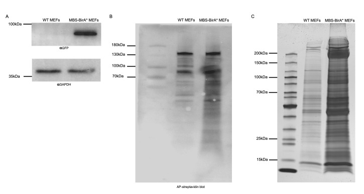
Figure 2. Functional validation of the integrated BirA* fusion construct.
A. Expression check of full-length BirA* construct via Western blot. The full-length fusion protein from the nls-HA-2x mcp-egfp-BirA* transgene, in total 91.4 KDa, is detectable by Western blot against the GFP part only in MBS MEF cells. GAPDH can be used as a negative control protein since it can be detected in lysates from WT and MBS-BirA* MEFs. B. Testing for BirA* activity in MEFs using AP coupled streptavidin. After incubation of MEFs with 50 μM biotin for 24 h, cells were lysed and 10 μg of total cell lysates from WT and MBS-BirA* MEF cells were loaded per lane. Biotinylated proteins were detected in a colorimetric assay by Western blot using AP-streptavidin. C. A pattern of biotinylated proteins in RNA-BioID obtained after purification via streptavidin-coupled magnetic beads. 1/10th of the amount of magnetic beads that are normally used for mass spectrometry analysis were boiled in 100 μl of SDS sample buffer. Twenty microliter of these were loaded on a 10% SDS-PAGE, separated and detected by silver staining. The number and intensity of protein bands from the lysate of MBS-BirA* MEFs are much larger than that from WT-MEFs, indicating successful RNA-dependent biotinylation and capturing of associated proteins.

