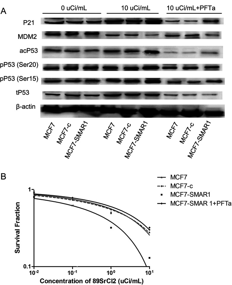
Figure 5.
p53 signaling pathway analysis by Western blot analysis (A) and clone formation assay (B). (A) Under the irradiation of 89SrCl2, the expression levels of pP53 (ser15), pP53 (ser20), acP53, and p21 in the MCF7-SMAR1 group were significantly increased, while the expression level of MDM2 was decreased compared with that in the MCF7 group or the MCF7-c group. However, after cells were treated with both 89SrCl2 and PFTα, the expression levels of p53 signaling pathway-related proteins in MCF7-SMAR1 group had no change compared with that in the MCF7 group or the MCF7-c group. (B) After cells were treated with both 89SrCl2 and PFTα, PFTα could reverse the SMAR1-enhanced radiosensitivity to 89SrCl2 in the MCF7-SMAR1 group.

