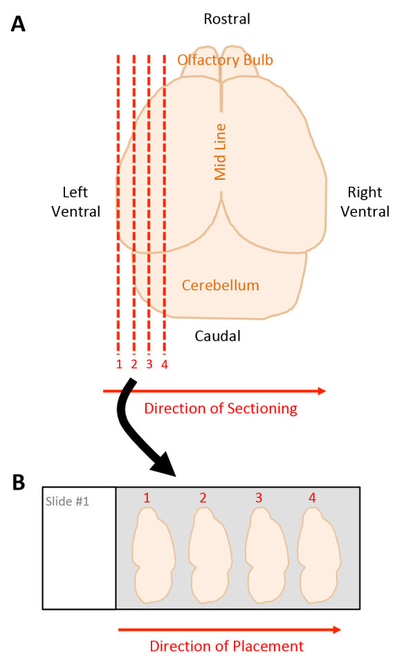
Figure 3. Schematic of tissue sectioning strategy.

A. Dorsal view of harvested mouse brain. Red dashed lines indicate sagittal sections beginning on the left ventral side moving towards the midline, starting with section 1 through 4. B. Placement of tissue sections onto microscope slide, moving left to right, starting with section 1 through 4.
