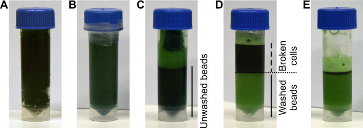
Figure 1. Breaking of Synechocystis cells.
A. Screwcap tubes are filled with 3 ml of glass beads and 3 ml of cyanobacterial suspension; B. The tubes are sealed with parafilm and cells lysed using a bead beater. C. After breaking cells, the beads are spun to the bottom and the supernatant is collected. D. The beads are washed with MES buffer; E. After each washing step, the supernatant is collected.

