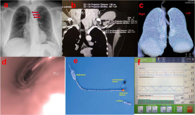
Figure 1.

(a) Chest X-ray showing tracheal deviation at the tbl4–T5 level. (b) & (c) Coronal cut contrast- enhanced MDCT showing narrowing at the tbl4–T5 level, along with an enlarged thyroid. (d) 3D Reconstruction of MDCT with virtual endoscopy showing tracheal narrowing. (e) Straw ETT (Tritube) used for the initial intubation. (f) CO2, intratracheal pressure, and tidal volume readings during the procedure.
