Figure 2.

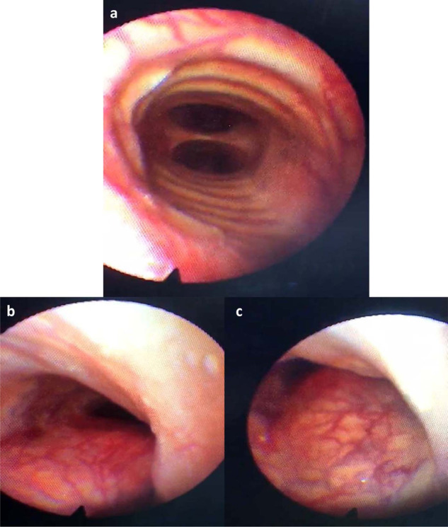
(a) shows the carina at the level below the stenosed area of the trachea, while (b) & (c) show the stenosed area in the trachea at the level of tracheal rings 5–6. The figures were obtained from fiber optic tracheoscopy.

(a) shows the carina at the level below the stenosed area of the trachea, while (b) & (c) show the stenosed area in the trachea at the level of tracheal rings 5–6. The figures were obtained from fiber optic tracheoscopy.