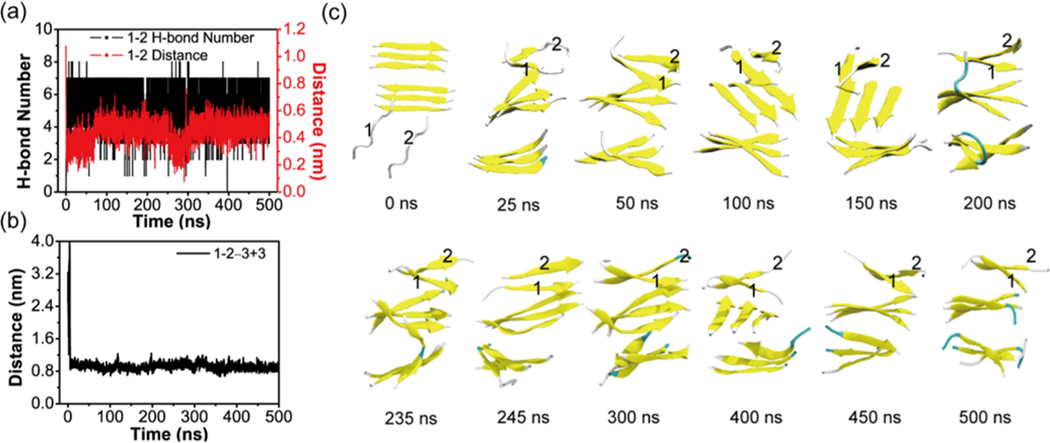
Figure 10.
Analysis of the MD trajectory on the system consisting of a 3 + 3 bilayer β-sheet and two random chains in conventional MD simulations. Time evolution of (a) the backbone H-bond number and the main-chain distance between chain 1 and chain 2. (b) The main chain center-of-mass distance between chain 1 + 2 and the upper layer β-sheet of the 3 + 3 bilayer β-sheet. (c) Representative snapshots at 12 different time points.

