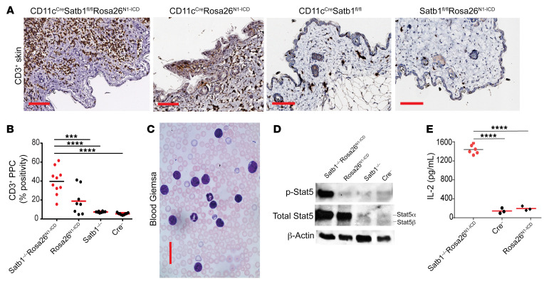
Figure 3. Satb1 ablation and Notch activation cooperate to transform postthymic CD8+ T lymphocytes into skin-homing lymphoma cells with phosphorylated Stat5 and cytokine increase.
(A) Accumulation of CD3+ T cells in skin of 10-week-old CD11cCreSatb1fl/flRosa26N1-ICD mice compared with single-genotype littermates. Scale bars: 100 μm. (B) Quantitative representation of positive pixel count (PPC) of CD3+ staining of skin from CD11cCreSatb1fl/flRosa26N1-ICD (n = 10), CD11cCreRosa26N1-ICD (n = 8), CD11cCreSatb1fl/fl (n = 8), and CD11cCre-negative mice (n = 8). One-way ANOVA with Tukey’s multiple-comparison test: ***P ≤ 0.001; ****P ≤ 0.0001. (C) Representative Giemsa staining of peripheral blood in the CD11cCreSatb1fl/flRosa26N1-ICD mice. Scale bar: 100 μm. (D) Western blot analysis of protein extracts from GFP+ (Notch1-overexpressing) cells sorted from the bone marrow of CD11cCreSatb1fl/flRosa26N1-ICD and CD11cCreRosa26N1-ICD mice, plus sorted CD8+ T cell splenocytes from wild-type (Cre–) and CD11cCreSatb1fl/fl mice. Representative of 2 independent experiments. (E) Immunopurified CD3+ T cells (2 × 10⁵) were stimulated with 0.5 μg/mL PMA and 1 μg/mL ionomycin in RMP1 with 10% FBS for 4 hours at 37°C. Supernatants were diluted 1:40 and IL-2 was quantified by ELISA (BioLegend) for CD11cCreSatb1fl/flRosa26N1-ICD (n = 6), CD11cCreRosa26N1-ICD (n = 3), and CD11cCre-negative mice (n = 3). One-way ANOVA with Tukey’s multiple-comparison test: ****P ≤ 0.0001.

