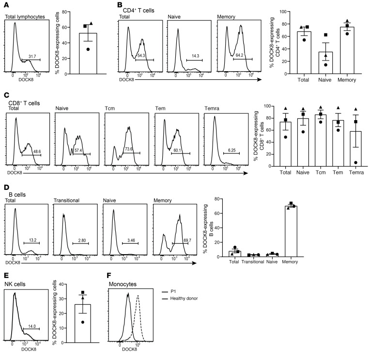
Figure 2. DOCK8-expressing cells are present in numerous lymphocyte populations and enriched in the memory compartments.
Patient PBMCs were stained for surface expression of CD4, CD8, CD20, CD56, CCR7, CD45RA, CD10, and CD27, fixed, permeabilized, and then stained intracellularly to detect DOCK8. Percentage of cells positive for DOCK8 expression in (A) total lymphocytes; (B) total, naive (CCR7+CD45RA+), and memory (CCR7±CD45RA–) CD4+ T cells; (C) total, naive, Tcm (CCR7+CD45RA–), Tem (CCR7–CD45RA–), and Temra (CCR7–CD45RA+) CD8+ T cells; (D) total, transitional (CD10+CD27–), naive (CD10–CD27–), and memory (CD10–CD27+) B cells; (E) CD3–CD56+ NK cells; and (F) monocytes were determined by flow cytometry. Circles, P1; squares, P2; triangles, P3. Solid line histograms depict P1 and dashed line histograms depict a healthy donor. Error bars represent SEM

