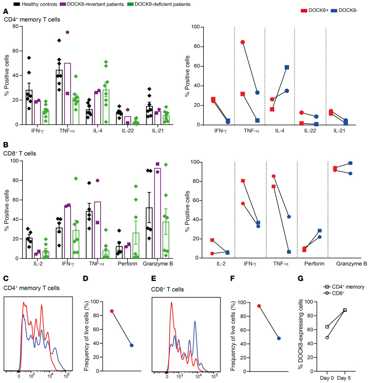
Figure 7. T cell function, proliferation, and survival is improved in DOCK8-revertant cells.
Sorted CD4+ memory T cells and CD8+ T cell from healthy donors (n = 5–9), DOCK8-revertant patients (circles, P1; squares, P2) and DOCK8-deficient patients (n = 7) were cultured for 5 days with TAE beads (anti-CD2, -CD3, -CD28). After restimulation with PMA/ionomycin, (A) CD4+ memory T cells were stained for intracellular expression of DOCK8 and the cytokines IFN-γ, TNF-α, IL-4, IL-22, and IL-21 in total cells and DOCK8+ and DOCK8– cells. (B) CD8+ T cells were stained for intracellular expression of DOCK8 and the cytokines IL-2, IFN-γ, and TNF-α, as well as perforin and granzyme B. (C–F) Proliferation (C, E) and survival (D, F) of DOCK8+ and DOCK8– CD4+ memory T cells and CD8+ T cells from P1 were determined by dilution of CFSE and staining with a live/dead marker. (G) The frequency of DOCK8-expressing cells in CD4+ memory T cells and CD8+ T cells before and after 5-day culture was determined in P1. Error bars are SEM.

