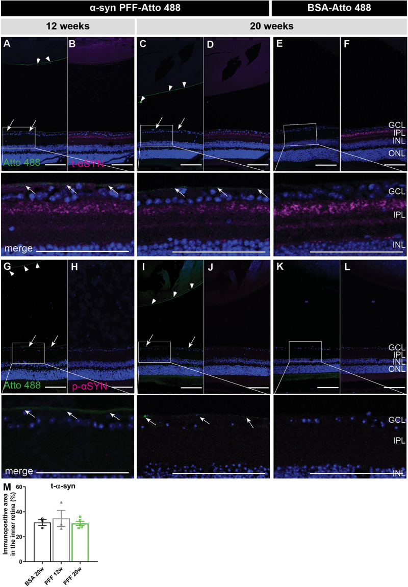
FIGURE 2.
Localization of α-syn in the eye at 12 and 20 weeks post IVT injection of α-syn PFFs. (A–D) Histological examination of sagittal eye sections demonstrates the absence of Atto 488-labeled α-syn PFFs in the retina at 12 weeks (A,B) and 20 weeks (C,D) post IVT injection. Limited Atto 488 signal is detected in the vitreous, predominantly at the ILM (arrows) and lens capsule (arrow heads). (E,F) Atto 488-labeled BSA is detected in retina nor vitreous at 20 weeks post injection. (A–F) Immunohistochemistry for total α-syn reveals presence of PFFs and endogenous α-syn, distributed similarly in the retina of eyes injected with α-syn PFF-Atto 488 and BSA-Atto 488. (G–L) Immunohistochemistry for p-α-syn shows its absence in the retina of eyes injected with α-syn PFF-Atto 488 and BSA-Atto 488. (M) Total α-syn in the retina was quantified by measuring the immunopositive area in the inner retina of animals injected with BSA (20 weeks) or α-syn PFFs (12 and 20 weeks post injection). Scalebar: 100 μm, n = 4. GCL, ganglion cell layer; IPL, inner plexiform layer; INL, inner nuclear layer; ONL, outer nuclear layer.

