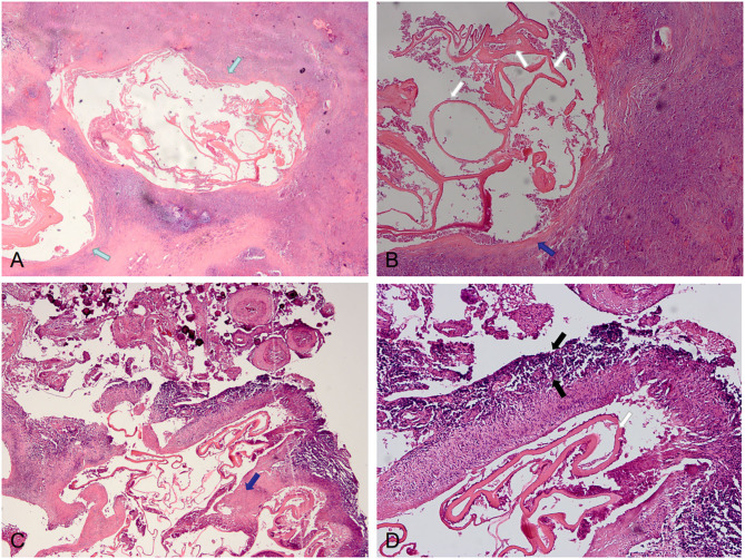
Figure 4.
Pathological features of IAE. Case 21: (A) Two lesions are visible in the parenchyma (green arrows), with obvious fibrosis around the lesions and unclear boundary with surrounding tissues. (B) Lesions with fibrotic cyst wall (blue arrow) and uniform red staining lamellar internal sacs (white arrows). Case 12: (C,D) There are fibrosis (blue arrows) of the cyst walls with calcification and inflammation (black arrows) at the periphery.

