Figure 5.
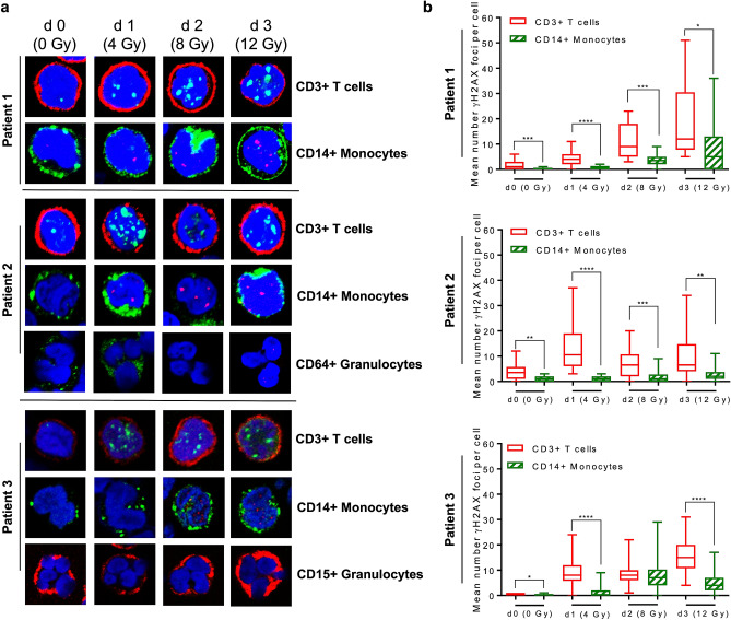
γH2AX staining in T cells (CD3+), monocytes (CD14+) and granulocytes (CD64+ or CD15+) isolated from patients during total-body radiation. (a) Representative images are shown of γH2AX foci (red or green) and CD surface marker (green or red) of PBMC at day 0 up to day 3 obtained from three different patients. Similar to what we observed for ex vivo irradiated granulocytes obtained from peripheral blood, no γH2AX foci could be detected in granulocytes upon exposure to ionising radiation in vivo. (b) Quantification of γH2AX foci in T cells and monocytes. In T cells significantly more γH2AX foci were induced following increasing cumulative doses of IR compared to monocytes. Depending on the yield and quality of the sample, 11 to 50 cells were counted per day and patient (Patient 1, 11 up to 20 cells; Patient 2, each data set 20 cells; Patient 3 each data set 50 cells). Box plots, t-test, *p < 0.05, **p < 0.01, ***p < 0.001, ****p < 0.0001.

