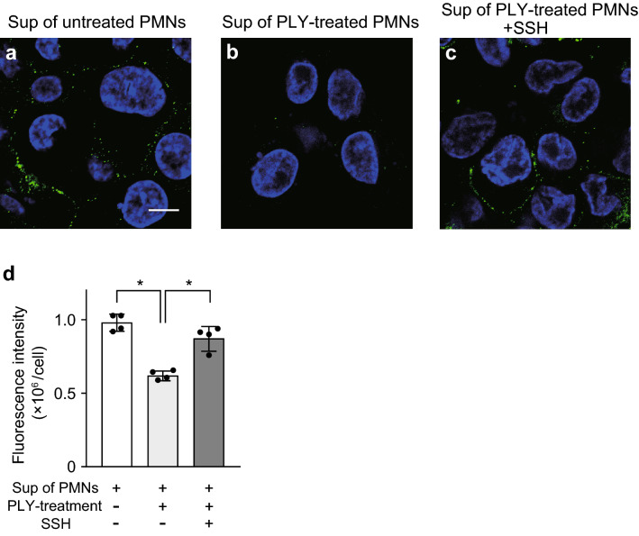
Figure 1.
Culture supernatant from neutrophils treated with pore-forming toxin decreases fluorescence intensity of HLA class II on macrophages. (a–c) THP-1-derived macrophages were exposed to culture supernatant (Sup) from untreated neutrophils (PMNs) or pneumolysin (PLY)-treated PMNs in the presence or absence of 100 μg/mL sivelestat sodium hydrate (neutrophil elastase inhibitor; SSH) for 6 h. Representative fluorescence microscopy images of macrophages stained for DNA (DAPI; blue) and HLA class II (HLA-DP β1; green) are shown. Scale bar: 10 µm. (d) Fluorescence intensity of HLA class II per cell was calculated. Data representing the mean ± SD of quadruplicate experiments was evaluated by one-way analysis of variance with Tukey’s multiple-comparisons test. *Significantly different between the indicated groups at P < 0.01.

