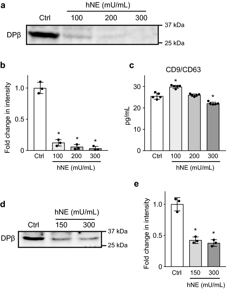
Figure 4.
Neutrophil elastase (NE) degrades HLA class II on extracellular vesicles derived from macrophages. (a) THP-1-derived macrophages were cultured in RPMI 1640 in the presence or absence of various concentrations of hNE (100–300 mU/mL) for 6 h. HLA-DP β1 expression on extracellular vesicles isolated from supernatant samples was determined by Western blot analysis. (b) Intensity of Western blotting signals of HLA-DP β1 on extracellular vesicles isolated from supernatant samples was quantified by densitometry. Data represent the means ± SD of triplicate experiments and were evaluated by one-way analysis of variance with Dunnett’s multiple-comparisons test. *Significantly different as compared with that of the control group at P < 0.01. (c) Extracellular vesicle production in supernatant samples were measured with a CD9/CD63 ELISA kit. Data represent the mean ± SD of quintuplicate experiments and were evaluated by one-way analysis of variance with Dunnett's multiple-comparison test. *Significantly different from control group at P < 0.01. (d) Extracellular vesicles were isolated from supernatant of THP-1 derived macrophages followed by hNE-treatment (150 and 300 mU/mL) for 4 h. Western blot analysis was performed to determine HLA-DP β1 expression in NE-treated and untreated extracellular vesicles. (e) Intensity of Western blotting signals of HLA-DP β1 on hNE-treated extracellular vesicles was quantified by densitometry. Data represent the means ± SD of triplicate experiments and were evaluated by one-way analysis of variance with Dunnett’s multiple-comparisons test. *Significantly different as compared with that of the control group at P < 0.01.

