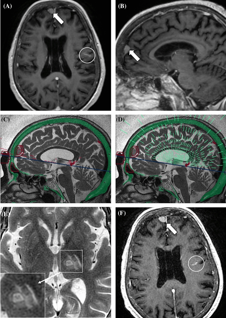
Figure 4.
81-year-old woman with essential tremor. Preoperative axial (A) and sagittal (B) multiplanar reconstructions (3 mm thick, average algorithm) from post-contrast T1w 3D BRAVO demonstrate a 11 mm extra-axial lesion compatible with meningioma (arrows). A developmental venous anomaly in the left fronto-parietal region is also present (white circle). Sagittal intraoperative T2-weighted FRFSE images show no-pass regions (red circles) with blocked [(C) red lines] and active [(D) green lines] elements. A total of 13 additional elements (11.6% of all blocked elements) were turned off due to the presence of the meningioma. 48-h follow-up axial T2-weighted FRFSE sequence (E) shows the FUS-placed lesion in the left nucleus ventralis intermedius. Post-contrast (F) image demonstrates no changes in the meningioma (arrow) or developmental venous anomaly (white circle).

