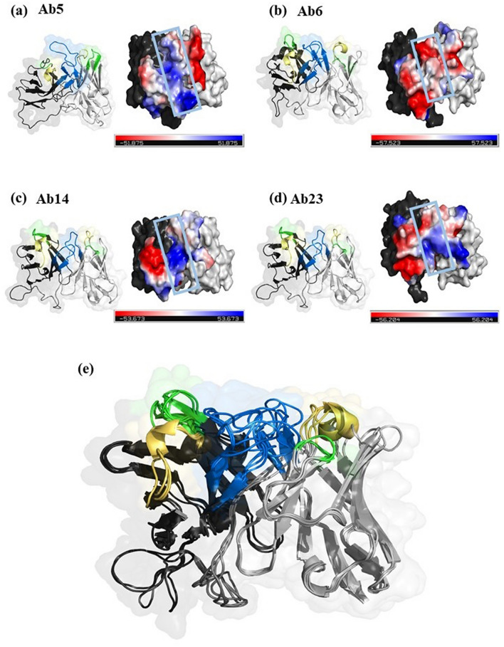
Figure 5.
Three dimensional structures of rNIE specific antibodies (a) Ab5, (b) Ab6, (c) Ab14 and (d) Ab23, by surface/ribbon (left) and electrostatic (right) presentations. Variable light (VL) and variable heavy (VH) domains are in black and grey ribbon presentation, respectively. CDR1, CDR2 and CDR3 are in yellow, green and blue ribbon presentation, respectively. Electronegative and electropositive residues are in red and blue surface presentations viewed from top. CDR3 is highlighted with blue box. (e) Superimposition of the four modelled scFvs. Figure was prepared by PyMol ver 2.4.0 (https://pymol.org/2/).

