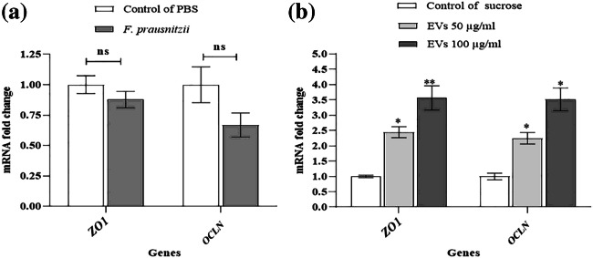
Fig. 3.
The effects of F. prausnitzii and its EVs on TJs: (a) the Caco-2 cells were treated with F. prausnitzii at MOI 10 and (b) different concentrations of F. prausnitzii–derived EVs (50 µg/ml and 100 µg/ml); *, ** p < 0.05 and p < 0.01 were considered statistically significant, respectively. ns represents no significance. GAPDH was used as an internal control

