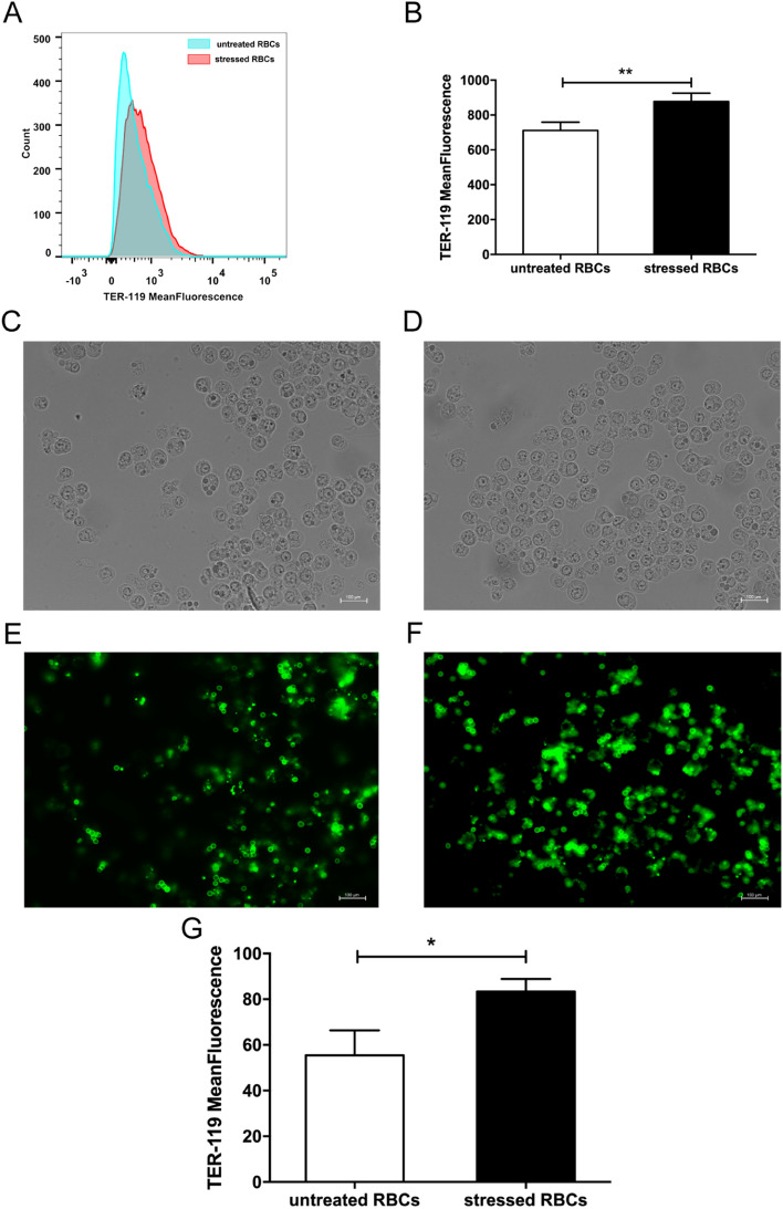
Figure 2.
Stressed RBCs tended to be phagocytosed. FITC-TER 119-labelled RBCs were incubated with BALB/c macrophage cell line Raw 264.7 (n = 9). (A,B) FACS results of erythrophagocytosis. Erythrophagocytosis was represented by FITC-mean fluorescence intensity. (C,F) Immunofluorescence results of erythrophagocytosis. Bright field showed macrophages treated with untreated RBCs (C) and stressed RBCs (D), fluorescence field showed TER-119 labelled untreated RBCs (E) and stressed RBCs (F). Arrow showed RBCs located in macrophages. (G) statistic results of erythrophagocytosis of untreated RBCs and stressed RBCs (*p < 0.05; **p < 0.01).

