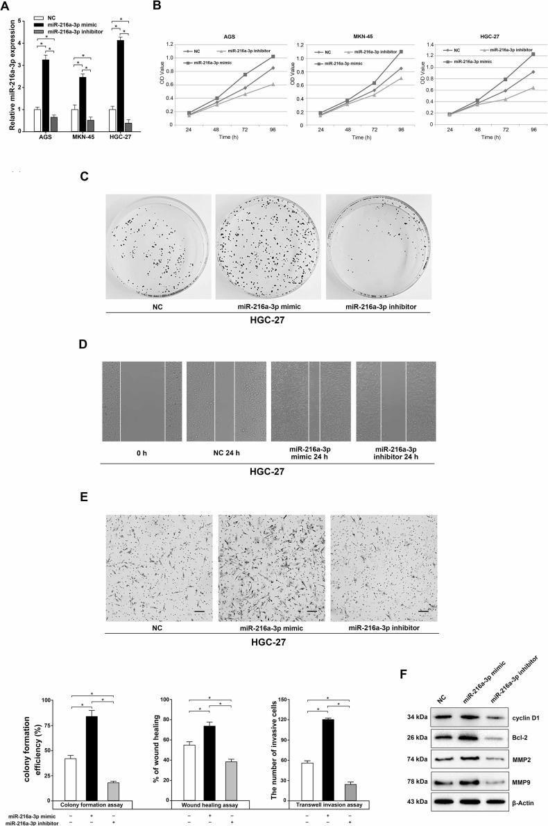
Figure 2.
Effects of miR-216a-3p on the proliferation, migration, and invasion of human GC cells. (A) The miR-216a-3p expression was measured by qRT-PCR based on whole-cell lysate from negative control (NC), miR-216a-3p mimic-, or inhibitor-transfected AGS, MKN-45, and HGC-27 cells. (B) MTS assay of proliferation in AGS, MKN-45, and HGC-27 cells following a time course of transfection with NC, miR-216a-3p mimic, or miR-216a-3p inhibitor. (C–E) The effects of miR-216a-3p on the plate colony formation efficiency (C), migration (D), and invasion (E) of human HGC-27 cells were assessed. The rate of wound healing was calculated with the following formula: (0-h width of wound − 24-h width of wound)/(0-h width of wound). (F) Effects of miR-216a-3p on cyclin D1, Bcl-2, matrix metalloproteinase 2 (MMP2), and MMP9 expression were measured by Western blot. All experiments were performed in triplicate. U6 or β-actin was used as the internal control. *p < 0.05, one-way analysis of variance (ANOVA).

