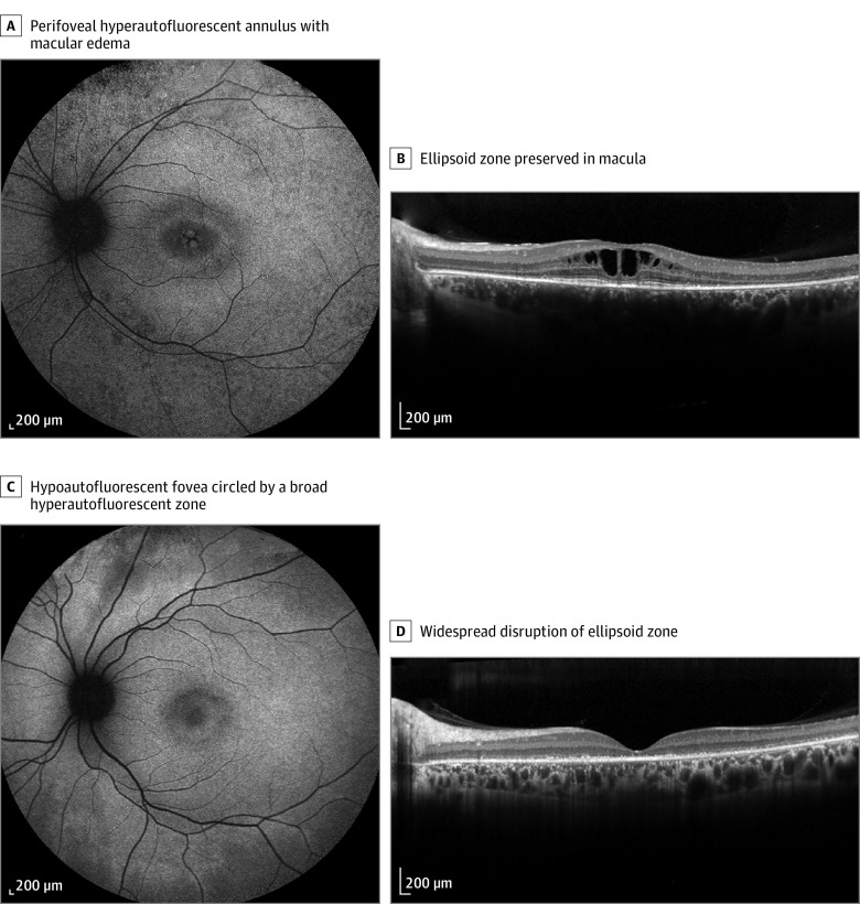
Figure 1. Example of Milder (A and B) and Severe (C and D) Forms of CLN3-Related Isolated Retinal Degeneration.
A, Short-wavelength fundus autofluorescence perifoveal hyperautofluorescent annulus with macular edema in patient CIC09853 with genotype M2, M8, and M11. B, Spectral-domain optical coherence tomography (horizontal scan) of the same patient revealed cystoid maculopathy with a preserved ellipsoid zone. C, Short-wavelength fundus autofluorescence in patient CIC09088 with genotype M5 and M9. D, Spectral-domain optical coherence tomography widespread disruption of ellipsoid zone in the same patient.

