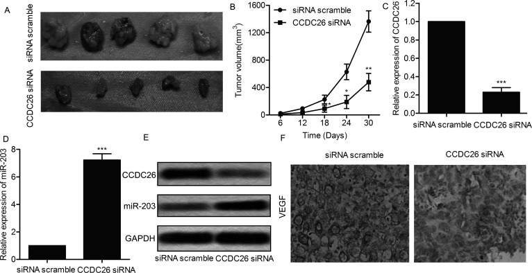
Figure 6.
CCDC26 depresses tumor growth and metastasis in vivo. U-251 cells were transfected with CCDC26-siRNA or siRNA scramble. A glioma xenograft mouse model was created by subcutaneous injection of U-251 cells to SPF nude mice. (A) Representative images of glioma xenograft tissues in each group (n = 5). (B) The diameter of glioma xenograft tissues in each group was measured every 6 days from tumor formation to 30 days. Tumor growth curve is shown as means ± SD. (C) Relative expression of CCDC26 in glioma xenograft tissues from each group was detected through qRT-PCR. (D) Relative expression of miR-203 in glioma xenograft tissues from each group was detected through qRT-PCR. (E) Expression of CCDC26 and miR-203 in glioma xenograft tissues from each group was detected through Northern blotting. GAPDH was used as an endogenous reference. (F) Expression of migration marker protein vascular endothelial cell growth factor (VEGF) in formalin-fixed, paraffin-embedded glioma tumors from each group was detected through IHC analysis. *p < 0.05, **p < 0.01, ***p < 0.001 versus the scramble group. The bars show means ± SD of three independent experiments.

