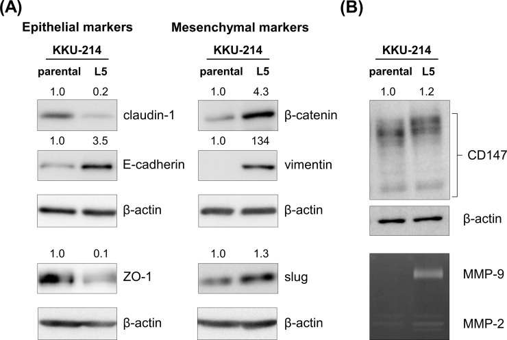
Figure 3.
Mesenchymal markers were upregulated in KKU-214L5 cells. Epithelial and mesenchymal markers were determined using Western blotting; β-actin was used as the loading control. (A) KKU-214L5 had downregulation of epithelial markers (claudin-1 and zona occludens 1 [ZO-1]) and upregulation of mesenchymal markers (β-catenin, vimentin, and slug), with an increase in epithelial (E)-cadherin. (B) Cluster of differentiation 147 (CD147) expression and matrix metalloproteinase 2 (MMP-2) and MMP-9 activities were elevated in KKU-214L5. Protein lysates of 5 μg were used for all epithelial–mesenchymal transition (EMT) markers, except ZO-1 and snail homolog 2 (slug) were at 30 μg. The numbers on the top of the Western blot indicate the relative protein band intensities by giving those of KKU-214 as 1. Parental = KKU-214; L5 = KKU-214L5.

