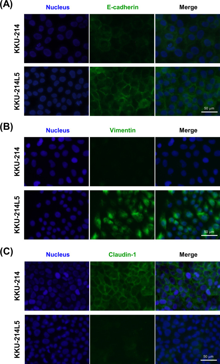
Figure 4.
Immunocytofluorescence staining of EMT markers in KKU-214L5 compared to KKU-214. The protein expression of E-cadherin, vimentin, and claudin-1 were determined using immunocytofluorescence stain with immunoglobulin G (IgG)-conjugated Alexa Fluor® 488 (green). Nuclei were stained with Hoechst 33342 (blue). Overexpression of (A) E-cadherin and (B) vimentin and suppression of (C) claudin-1 was observed in KKU-214L5 cells. Scale bar: 50 μm.

