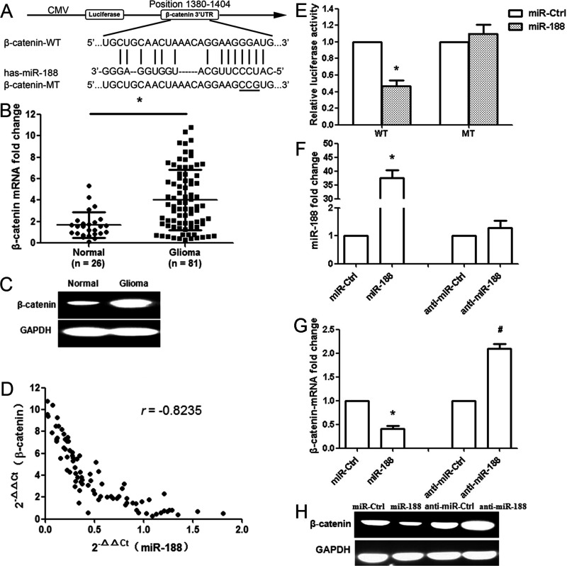
Figure 2.
miR-188 directly targets the β-catenin gene. (A) Bioinformatics predicted interactions of miR-188 and their binding sites at the 3′-untranslated region (3′-UTR) of β-catenin (PicTar and miRanda). CMV, cytomegalovirus; MT, mutant; WT, wild type. (B) β-Catenin mRNA expression in glioma (n = 81) and normal brain tissues (n = 26). (C) β-Catenin protein level was measured by Western blot. Relative optical density was normalized to glyceralde 3-phosphate dehydrogenase (GAPDH) to indicate protein content. (D) miR-188 and β-catenin levels were inversely correlated. 2−ΔΔCt values of miR-188 and β-catenin were subjected to a Pearson’s correlation analysis (n = 81, r = −0.8235, p < 0.001, Pearson’s correlation). (E) The luciferase reporter plasmid containing wild- or mutant-type β-catenin 3′-UTR was cotransfected into HEK293T cells in combination with miR-188 or miR-Ctrl. Luciferase activity was examined by the dual-luciferase assay. (F) The miR-188 expression was determined in glioma LN229 cells after miR-188 overexpression or anti-miR-188 treatment. (G) The β-catenin mRNA was determined after miR-188 overexpression or anti-miR-188 treatment. (H) The expression of β-catenin protein was analyzed by Western blot. GAPDH was used as control. *p < 0.01, compared with the miR-Ctrl group; #p < 0.01, compared with the anti-miR-Ctrl group, n = 3.

