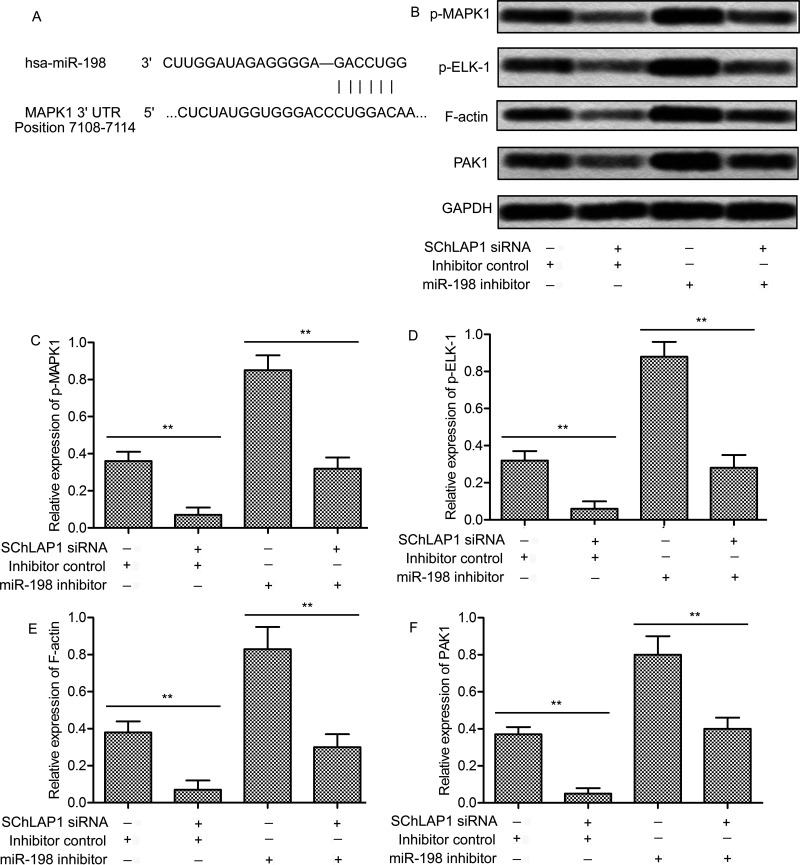
Figure 6.
SChLAP1 activates the MAPK1 signaling pathway. (A) Bioinformatics analysis of miR-198 and MAPK1. (B) PC-3 cells were transfected with SChLAP1-siRNA and/or the miR-198 inhibitor for 24 h, and the levels of phosphorylated MAPK1, phosphorylated ELK-1, F-actin, and PAK1 were measured by Western blot. (C–F) Quantification of Figure 3B. All experiments were repeated at least three times. GAPDH was used as a loading control. **p < 0.01.

