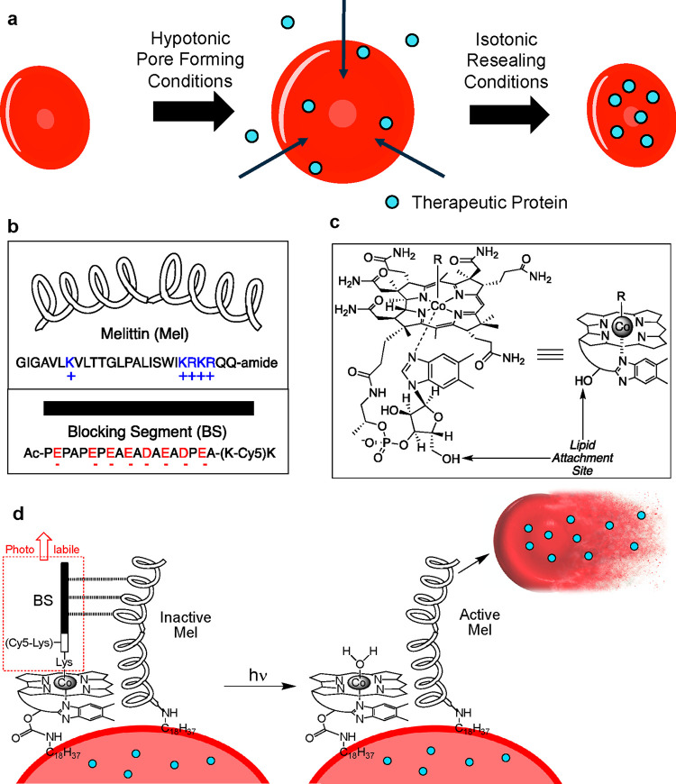
Figure 1.
Assembly of a photohemolysis trigger on the surface of RBCs. a. Both human and mouse RBCs are first internally loaded with a therapeutic protein (blue circles) by sequential treatment with hypotonic conditions (pore formation), the protein of interest, and isotonic buffer (pore resealing). b. The photolytic trigger is composed of two peptides, melittin (Mel) and its pro-domain blocking segment (BS). c. Cobalamin (Cbl) is synthetically modified with a lipid, and the BS peptide is appended as a photocleavable ligand to Co. d. Simultaneous exposure of RBCs to the lipidated BS and Mel peptides generates a photoresponsive RBC construct (left). Upon illumination (525 nm for C18-Cbl-BS or 660 nm for C18-Cbl-Cy5BS), the BS peptide is released from Cbl, generating active Mel, subsequent hemolysis, and release of the internally loaded protein therapeutic. C18-Cbl-Cy5BS (Scheme S4) is an analog of C18-Cbl-BS that responds to 660 nm exposure.

