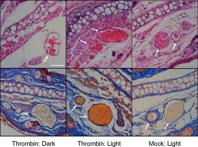
Figure 5.

Murine RBCs internally loaded with buffer or thrombin and surface modified with C18-Mel/C18-Cbl-BS were tail vein injected into healthy FVB mice (n = 4 for each experimental condition). A 1 mm2 region of one ear from each mouse was illuminated (561 nm) under a confocal microscope, the mice were euthanized, and both “dark” and “light” ears were harvested. Four μm cross sections of the fixed tissues were stained with H&E (top row) and Martius Scarlet Blue dyes (bottom row). Left: “Dark” ear reveals healthy blood vessels with RBCs stained yellow (bottom image). Middle: “Light” ear displays the presence of fibrin (orange/red; bottom image) and significant vascular congestion. Right: Light exposed ear from animals injected with photoresponsive RBCs that were internally loaded with buffer. The blood vessels do not display fibrin or venous congestion as evidenced by the free space between the RBCs and the endothelium. Scale bar = 50 μm.
