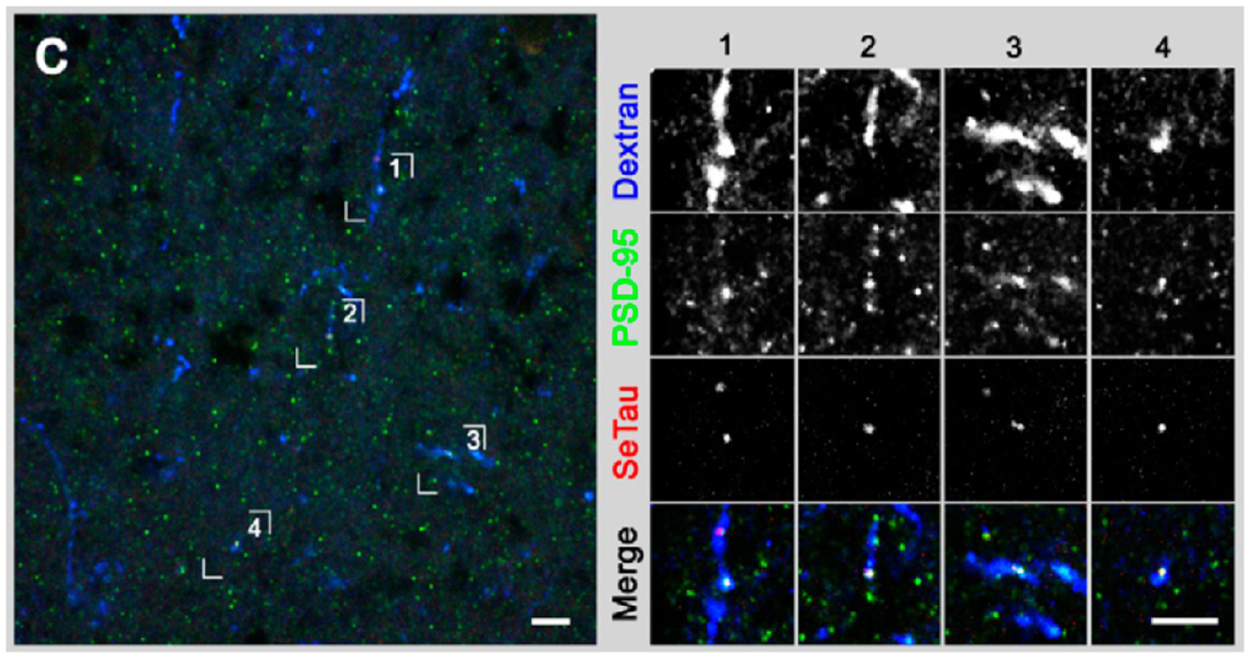
Figure 10.

Colabeling of tadpole brain with SeTau-peptide conjugate (red) and fluorescent anti-PSD-95 antibody (green) (left). In the expanded views of the highlighted regions (right), the SeTau conjugate shows strong colocalization in the neurons stained with anti-PSD-95 antibody. Cascade blue dextran was used as a space filler. Image used with permission from ref 48. Copyright 2012 Podgorski et al.
