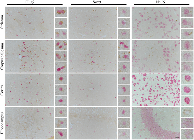
Figure 4.
The cell- and side-specific expression of four repeats of tau (4R) isoform in hTau animals brain. The high-power microphotography of double immunohistochemistry staining of 4R tau isoform (RD4, brown chromogen) combined with oligodendrocytes (Olig2, left panel, red/pink chromogen), astrocytic (Sox9, middle panel, red/pink chromogen) or neuronal (NeuN, right panel, red/pink chromogen) markers. The representative pictures of four brain regions: striatum (upper panel), corpus callosum (second upper panel), cortex (third upper panel), and hippocampus (lower panel) with higher magnification of representative cells on the right side of each image. In striatum and corpus callosum only oligodendrocytes were RD4 positive, and no RD4 immunoreactive astrocytes or neurons were detected. In cortex only, some oligodendrocytes and rear neurons were RD4 positive, and no RD4 immunoreactive astrocytes were identified. Similarly, in the hippocampus, no astrocytes were RD4 positive, and only some oligodendrocytes and neurons were weakly RD4 immunoreactive. The scale bar represents 25 μm and applies to all pictures; inserts represent 4× digital enlargement.

