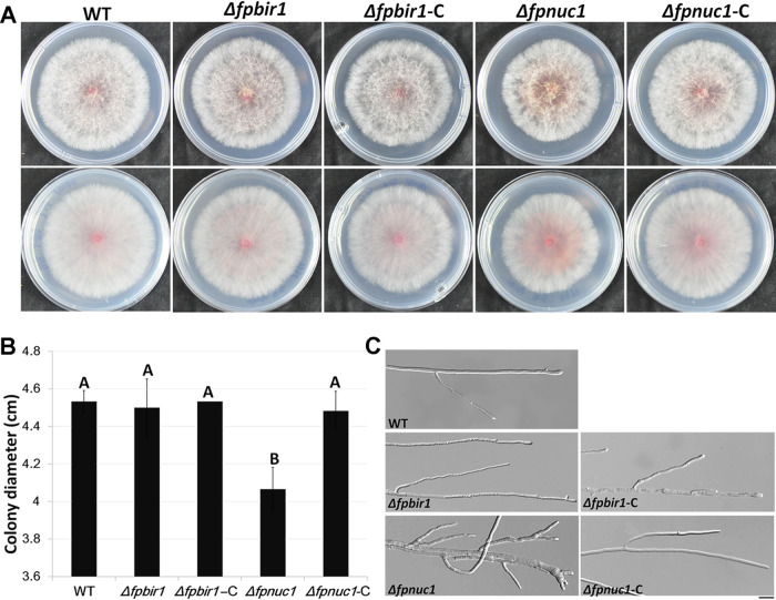
FIG 5.
Colony morphology and hyphal growth. (A and B) Colonies of all five strains grown on PDA medium. Photographs and colony diameters were taken after 3 days. The data shown are representative of those from three separate experiments. A and B above bars indicate significant differences at a P value of <0.01 using Duncan’s multiple-range analysis. (C) Hyphae of all five strains cultured on PDA plates for 24 h. Bar = 20 μm.

