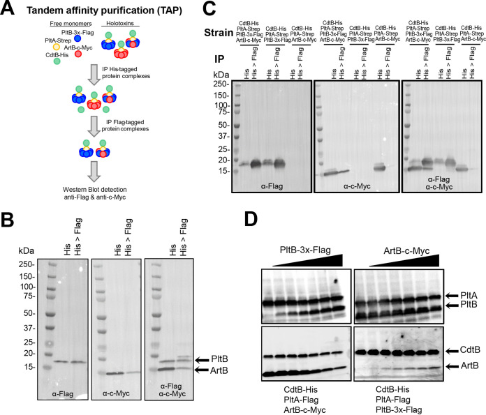
FIG 4.
PltB replaces ArtB for inclusion in the holotoxin. (A) Schematic for the approach used for TAP and Western blot detection of heteropentameric binding subunits. (B) Detection of PltB-3×-Flag and ArtB-c-Myc using TAP and Western blotting. Holotoxins were pulled down from E. coli BTH101 strains expressing CdtB-His, PltA-Strep, PltB-3×-Flag, and ArtB-c-Myc using α-His antibodies (His) followed by purification of PltB-3×-Flag-containing holotoxins by pulling down with α-Flag magnetic beads (lane His>Flag). Proteins were visualized with antibody staining to detect PltB-3×-Flag (left), ArtB-c-Myc (middle), and both PltB-3×-Flag and ArtB-c-Myc (right). Results of additional replicates can be found in Fig. S2 in the supplemental material. (C) Immunoblots demonstrating the specificity of the pulldown assays and antibody detection used for TAP (additional blots for additional replicates can be found in Fig. S2). (D) Results of the binding subunit exchange assay. PltB-3×-Flag was added at various concentrations to lysates containing CdtB-His, PltA-Flag, and ArtB-c-Myc (left), or ArtB-c-Myc was added to lysates containing CdtB-His, PltA-Flag, and PltB-3×-Flag (right). Holotoxins were purified by pulling down with α-His antibodies to detect CdtB-His, and subunits were detected using tag-specific antibodies (PltA-Flag, CdtB-His, and PltB-3×-Flag). Results from one representative experiment are shown; the assay was performed as three independent experiments. Images for additional replicates can be found in Fig. S2.

