FIG 1.
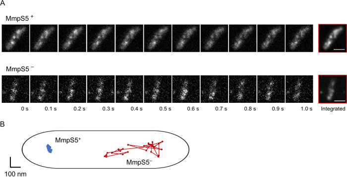
Dynamics of GFP-labeled MmpL5 in the inner membrane. (A) Real-time observation of MmpL5-GFP foci using TIRF microscopy. The BCG_0727-mmpS5-mmpL5 deleted strains were complemented by pKRB32 or pKRB29. These cell images present the dynamics of the fluorescent foci of MmpL5-GFP in the presence (top) or absence (bottom) of MmpS5. Data were acquired at 33 ms per frame. Bars, 1 µm. (B) The x-y trajectories of MmpL5-GFP in the presence (blue line) or absence (red line) of MmpS5.

