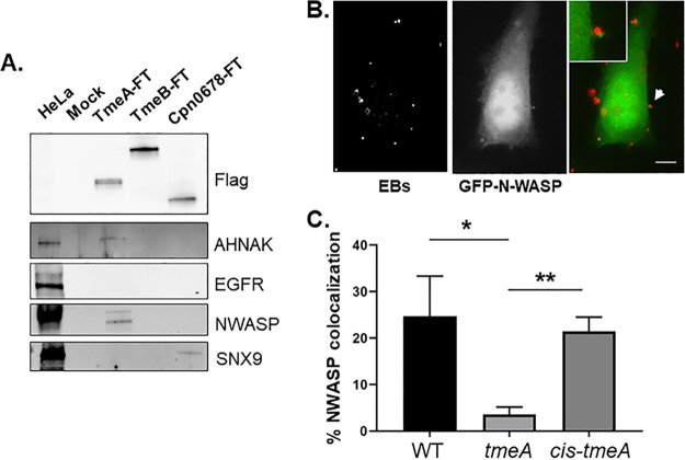
FIG 3.
TmeA interacts with N-WASP, and N-WASP recruitment to sites of invading EBs requires TmeA. (A) Flag-tagged proteins were immunoprecipitated from whole-cell lysates of HeLa cells expressing TmeA-FT, TmeB-FT, or Cpn0678-FT for 24 h. Immunoprecipitation from mock-treated lysates served as a negative control. Eluted material was probed in immunoblots for tagged chlamydial proteins using anti-Flag antibodies. Host proteins were detected using antigen-specific antibodies, and HeLa whole-cell lysates were included as a positive control for these antibodies. (B). GFP-N-WASP (green)-expressing HeLa cells were infected for 10 min with WT C. trachomatis (red) and visualized by epifluorescence microscopy. The arrow indicates the field of view shown as an inset in the merged image. Bar = 5 µm. (C). HeLa cells were cultivated for 20 min after synchronous infection (in triplicate) with WT, ΔtmeA, or cis-tmeA strains. Monolayers were stained for NWASP and Chlamydia using specific antibodies, and the percentage of colocalization was enumerated for ca. 100 randomly selected EBs. Data are represented as the percentage of EBs exhibiting adjacent NWASP localization. Statistical significance was computed using Student’s t test with Welch’s correction (*, P < 0.04; **, P < 0.002).

