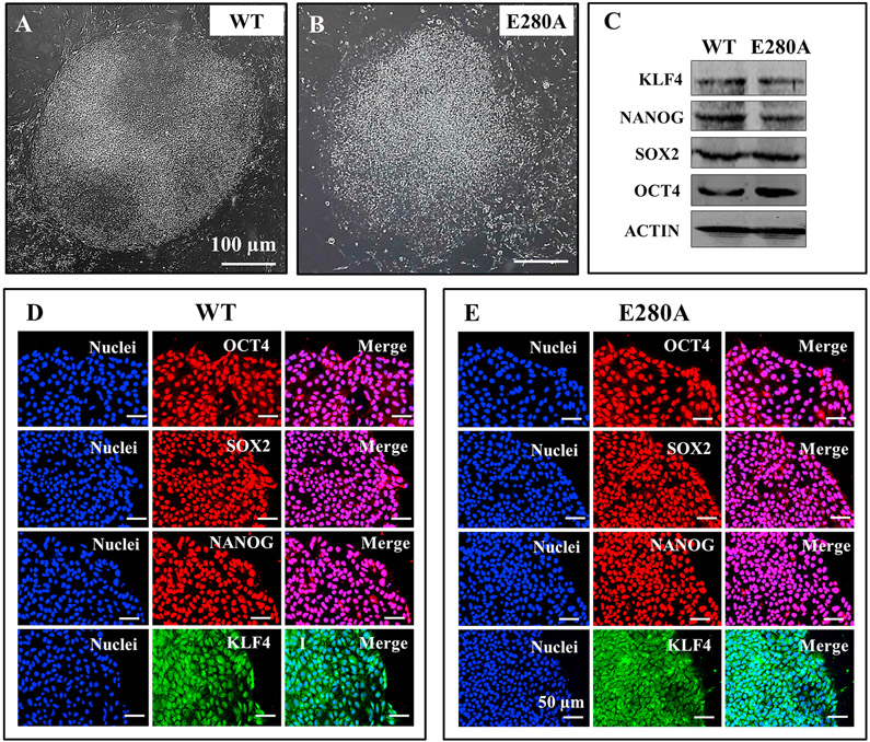
Fig. 1. Characterization of wild type and PSEN 1 E280A iPSCs.
(A) Representative light microscopy image of iPSCs colonies formed from a healthy individual; (B) Representative light microscopy image of iPSCs colonies formed from an AD patient carrying PSEN1 E280 A mutation. (C) Representative western blot images of the pluripotency markers KLF4, NANOG, SOX2 and OCT4 in iPSCs; (D–E) Representative immunocytochemistry images of iPSCs stained for OCT4, SOX2, NANOG, and KLF4 in WT (D) and PSEN 1 E280 A mutation (E). Nuclei are stained with Hoechst (blue). Scale bars 50 μm.

