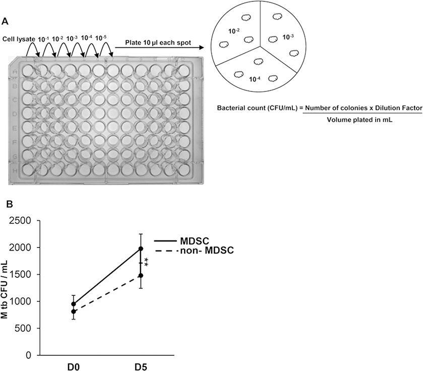
Fig. 2.
Intracellular replication of M. tuberculosis in MDSC isolated from peripheral blood mononuclear cells. (a) Sorted MDSC and non-MDSC are infected with M. tuberculosis Erdman at multiplicity of infection of 5 for 3 h, washed with D-PBS to remove non-phagocytosed bacteria and loosely adherent cells. Cells are treated with gentamicin (50 μg/mL) for 1–2 h at 37 °C and 5% CO2 to kill extracellular bacteria. Cells are lysed with 0.07% SDS and cellular lysates are serially diluted and plated in triplicate on Middlebrook 7H10 agar supplemented with OADC enrichment. The number of colonies are counted after 3 weeks and colony forming units (CFU)/ml determined. (b) Intracellular growth of M. tuberculosis shown at days-0 and −5 postinfection of MDSC and non-MDSC isolated from HIV-infected individuals. Data show mean values ±SEM; N = 4 donors. **p < 0.005

