Figure S2.
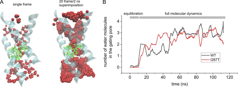
Water accessibility of the gating pore in WT and I287T mutant voltage sensor domain. (A) Left: 3-D structure of the Shaker VSD carrying the mutation I287T, obtained after 100 ns of a MD simulation in which water was allowed to equilibrate inside the intracellular and extracellular vestibules. The protein backbone is represented in cyan/transparent, the side chains of the gating pore residues are shown in licorice representation (green), and water molecules are shown in van der Waals representation (red). Right: Same as the structure shown to the left, but here, the water molecules from 20 consecutive frames of the simulation (one every 0.1 ns) are superimposed in order to define the WA region of the VSD. Note that only a very short region inside the gating pore is totally inaccessible to water. (B) Plot of the mean number of water molecules inside the gating pore region (defined as the water molecules residing inside a cylinder of 5 Å radius, extending from −1 Å to 6 Å with respect to the z component of the center of mass of the F290 aromatic ring) as a function of the simulation time. The black and red lines refer to the simulation performed with the WT structure (the same shown in Fig. 2) and with the mutant (I287T) structure, respectively.

