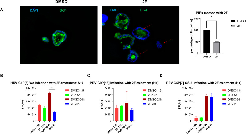
Fig 6. PIEs treated with 2F.
500 μM 2F or DMSO was added to differentiated PIEs for 3 days (refreshed medium and added new 2F or DMSO every day). Then 2F-treated PIEs were used either for A) IF detection of H antigen expression (BG4 antibody). BG4 (green) and DAPI (blue) staining are shown in a merged picture to denote the membrane staining pattern of H antigen. Left panel: H expression under DMSO treatment. Middle panel: H expression under 2F treatment. Red arrows denote PIEs with reduced H antigen expression. Right panel: Quantitive data for IF results. H+ and total cell numbers were counted and percentage of H+ cell s(%) are illustrated.

