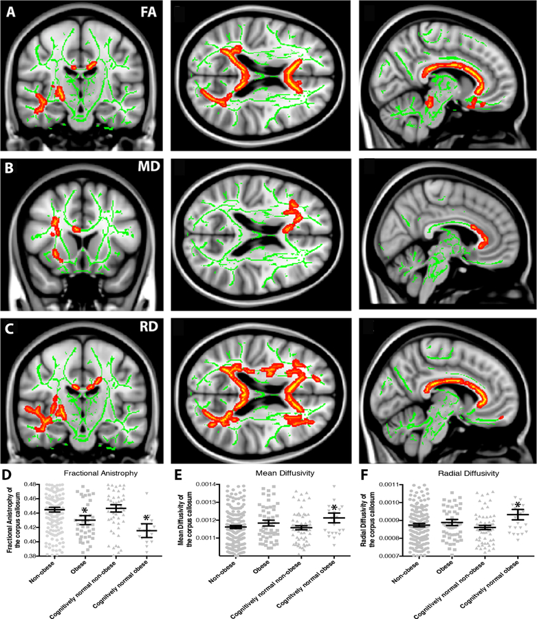
Figure 4. White matter integrity is decreased in obese individuals.
Cognitively normal older adults (CN) and patients with mild cognitive impairment (MCI) and Alzheimer’s disease (AD) who are obese (BMI≥30) showed reduced white matter integrity relative to non-obese (BMI<30) individuals. (A, B) Reductions in fractional anisotropy (FA), a general measure of white matter integrity, were observed in obese (n=54) relative to non-obese (n=202) individuals throughout the corpus callosum and frontal white matter. (B) On a regional basis, mean FA in the corpus callosum was reduced in obese relative to non-obese individuals both in the full sample and in CN older adults only (n=88, *p<0.05). (C, D) Increased mean diffusivity (MD), a reflection of increased white matter damage, was also observed in the frontal white matter in obese relative to non-obese individuals. (D) Regional analysis shows a significant increase in MD in the corpus callosum only in CN individuals (*p<0.05). (E, F) Increased radial diffusivity (RD), reflecting either demyelination or axonal swelling, was also observed in obese relative to non-obese individuals in the corpus callosum and frontal white matter. (F) Upon regional analysis, mean corpus callosum RD was increased in obese relative to non-obese individuals in the CN only group (*p<0.05).

