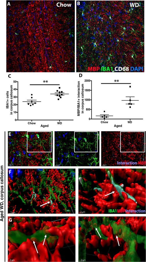
Figure 6. WD causes activation of phagocytosing myeloid cells.
(A, B) A representative image of the corpus callosum from an aged WD mouse showing myelin (MBP), myeloid cells (IBA1) and phagosome-containing myeloid cells (CD68). (C) There was a significant increase of IBA1+ cells in the corpus callosum of aged WD compared to aged chow mice (n≥7, **p= 0.0012). (D) Myelin-myeloid cell interactions were also significantly increased in aged WD compared to aged chow mice (n≥5, **p= 0.0035). (E–G) IMARIS was used to identify myeloid cells that were actively phagocytosing myelin. The image shows activated myeloid cells interacting with myelin visualized with anti-MBP (E). F and G are higher resolution images from the boxed region in E and show MBP (red) contacting myeloid cells (green). Labeling in purple shows the interactions between CD68+IBA1+ cells. Arrows (F, G) show MBP inside the cell body of the myeloid cells.

