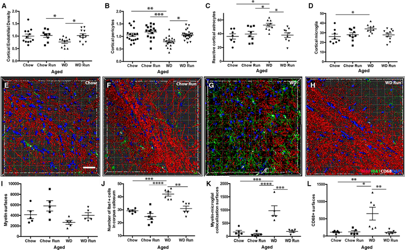
Figure 8. Running prevents WD-induced cerebrovascular damage and increases in phagocytosing myeloid cells.
(A-D) Running prevented WD-induced endothelial cell loss (A, n≥11, chow run vs. sedentary WD *p=0.049, sedentary WD vs. running WD *p=0.040) and the associated decrease in the number of PDGFRβ+ pericytes covering the blood vessels (B, n≥11, sedentary chow vs. sedentary WD **p=0.005, running chow vs. sedentary WD ***p=0.0007, sedentary WD vs. running WD *p=0.02). Running also prevented the increase in GFAP+ astrocytes surrounding blood vessels(C, n≥9, sedentary chow vs. sedentary WD *p=0.011, running chow vs. sedentary WD *p=0.047, sedentary WD vs. running WD *p=0.018) and the increase in IBA1+ myeloid cells (D, n≥9, running chow vs. sedentary WD *p=0.026). (E-H) Representative 3D reconstructions of the corpus callosum using IMARIS software of chow sedentary (E), chow run (F), WD sedentary (G) and WD run (H). Images show myelin (MBP, red), DAPI (blue), myeloid cells (IBA1, green) and CD68 (white) to identify phagocytosing myeloid cells. (I-L) Aged running WD mice tended to have more myelin surfaces than aged sedentary WD mice (I). However, in the corpus callosum, running prevented the WD-dependent increase in IBA1+ myeloid cells (J, sedentary chow vs. sedentary WD ***p=0.0002, running chow vs. sedentary WD ****p<0.0001, sedentary WD vs. running WD **p=0.001), the increase in myelin-myeloid cell interactions (K, sedentary chow vs. sedentary WD ***p=0.0002, running chow vs. sedentary WD ****p<0.0001, sedentary WD vs. running WD ***p=0.0003) and the increase in CD68+ surfaces (L, sedentary chow vs. sedentary WD **p=0.01, running chow vs. sedentary WD *p=0.011, sedentary WD vs. running WD **p=0.006). Scale bar for all images 40µm.

