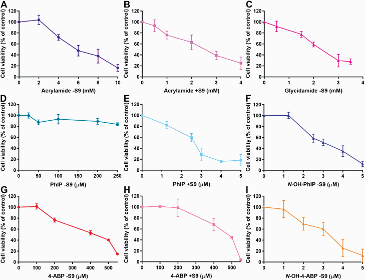
Fig. 1.
Cell viability assessment in FE1 cells. Cells were treated with various concentrations of acrylamide −S9 (A), acrylamide +S9 (B), glycidamide −S9 (C), PhIP −S9 (D), PhIP +S9 (E), N-OH-PhIP −S9 (F), 4-ABP −S9 (G), 4-ABP +S9 (H) or N-OH-4-ABP −S9 (I) for 6 h followed by a 72-h sampling time. Cell viability (% control) was assessed by staining with crystal violet. Cells treated with water (A–C) or DMSO (D–I) served as controls. Shown are mean values ± SD (n > 3).

