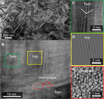
Fig. 2. Deformation microstructure of the swaged CrMnFeCoNi HEA subjected to quasi-static compression.

(A) TEM bright-field image shows profuse planar deformation features. (B) High-resolution TEM micrograph of the heavily deformed area with three distinctive regions of deformation twinning, hcp phase, and an amorphous region. The corresponding lattice images are given in (C to E), respectively. The hcp region in (D) is Fourier transformation–filtered to maximum the phase contrast, as detailed in the Supplementary Materials. The amorphous island, in (B), is formed at the intersection of hcp and twin bands.
