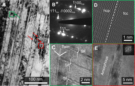
Fig. 3. Deformation microstructure of the CrMnFeCoNi HEA subjected to dynamic compression/shear.

(A) Bright-field image of the twins, stacking faults, hcp phase, and amorphous bands in the vicinity of the macroscopic adiabatic shear band, as schematically shown in Fig. 1E. (B) Selected-area electron diffraction pattern shows the existence of the fcc matrix, twinning spot, and the hcp reflections. (C) High-resolution TEM image shows the coexistence of nanoscale twin and the stacking faults (SF). (D) Fourier-filtered lattice image of the interface between hcp and fcc phases. (E) High-resolution TEM image of the amorphous bands [red square in (A)] and the corresponding fast Fourier–transformed diffractograph.
