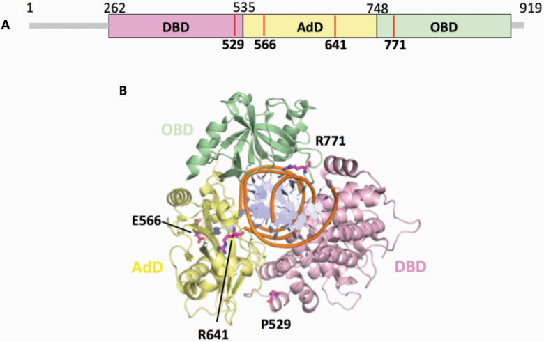
Figure 5.
Domain architecture of LIG1 with the disease mutations. (A) Schematic view showing the domain composition of human LIG1, including the N-terminal domain (amino acids 1–261, gray), and the catalytic core (amino acids 262–919) consisting of the DBD (pink), AdD (yellow) and OBD (green). (B) LIG1 (cartoon) in complex with an adenylated nicked DNA complex (stick, orange). The amino acid residues (magenta), P529, E566, R641 and R771, that are mutated in LIG1-deficiency disease are shown as sticks.

