
Figure 2.
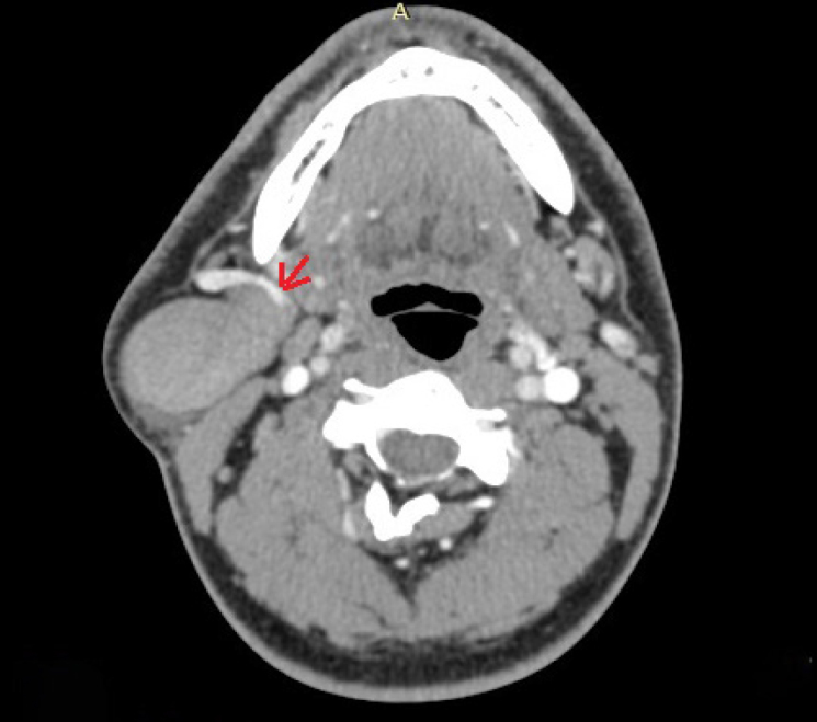
Axial images of the contrast enhanced computed tomography of the neck, showing venous ectasia of retromandibular vein (red arrow)
Official websites use .gov
A
.gov website belongs to an official
government organization in the United States.
Secure .gov websites use HTTPS
A lock (
) or https:// means you've safely
connected to the .gov website. Share sensitive
information only on official, secure websites.
Axial images of the contrast enhanced computed tomography of the neck, showing venous ectasia of retromandibular vein (red arrow)