
Figure 1.
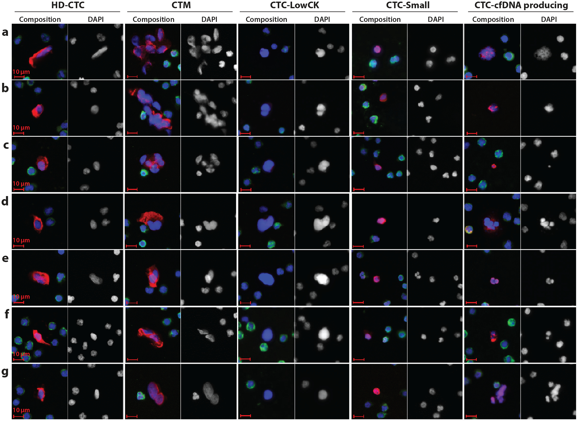
Candidate cells of circulating tumor cell (CTC) detection by the high-definition single-cell analysis (HD-SCA) approach of Kuhn and colleagues (J.-A. Thiele, P. Pitule, P. Ostašov, V. Liška, M. Králíčková, et al., unpublished data). The images represent the pleomorphic population of candidate cells found in the blood (before surgery) of metastatic colorectal cancer patients; these cells are stained with DAPI (blue), CK-pan mix (red), and CD45 (green). The composite image and the DAPI image are displayed for each cell type. Cell type abbreviations (from left to right): CTCs detected by the HD-SCA platform (HD-CTCs), CK-positive and CD45-negative cells with a nucleus distinct from WBC nuclei; circulating tumor microemboli (CTM), HD-CTC clusters (at least two or more HD-CTCs); CTC-LowCK, cells with a nuclear shape different from that of WBCs but CK negative and CD45 negative; CTC-Small, CK-positive and CD45-negative cells with a small (WBC-like) nucleus; CTC-cfDNA producing, CTCs undergoing apoptosis with irregular nuclear or cytoplasmic condensation and a possible source of circulating tumor DNA.