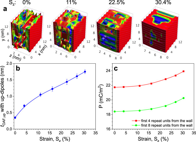
Fig. 5. Computer simulation of the direct piezoelectricity in PVDF.
a The color-coded simulation slab for PVDF after equilibration at 300 K for 2 ns. The color scale represents PVDF units with positive dipole moments along the y-axis with values between zero (blue color) and 2.1 D (red color). The empty space in the pictures is comprised of negative dipoles. There are 12 chains along the x-axis, 12 chains along the y-axis, and 30 repeat units along the z-axis. The slab thickness along z is 6.8 nm. Chain ends are attached to both slab walls with a rigid C–C bond (dipole moment fixed along the y-axis). The attachment points are organized in the same manner as the β crystal. The ab unit cell dimensions on the slab wall are a = 2.1 nm and b = 1.2 nm, respectively, which are about twice larger than the actual unit cell dimensions of the β crystal. The strain along z (Sz) is 0%, 11%, 22.5%, and 30.4%, respectively. b The tOAF,up and c polarization along y (Py) for the first 4 and 8 repeat units from the wall as a function of Sz.

