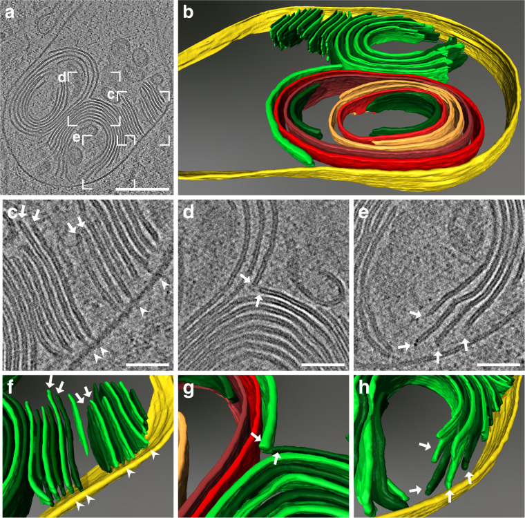
Fig. 4. Cryo-ET of LB-like organelles reveals structural details of membrane sheet termini and “T”-junctions.
a Central slice of the reconstructed tomogram acquired at the site of the correlated ABCA3-eGFP signal (correlation shown in Supplementary Fig. 7a, b). b Manual rendering of the tomogram (a). The limiting membrane of the LB is labeled in yellow, membrane sheets are labeled in green. To distinguish individual membrane sheets, alternating shades of green were used. Continuous membranes are labeled in red. A spiral-coiled membrane sheet is labeled in orange. c–e Detailed views of the reconstructed tomogram. Perpendicularly oriented membrane sheets toward the limiting membrane are often connected via a thin density (“T”-junction) to the limiting membrane of the LB (arrowheads). Membrane sheets show a rounded density at the membrane termini (arrows). f–h 3D rendering of detailed views (c–e). This figure is visualized as Supplementary Movies 2 and 3. Scale bars: a 200 nm, c–e 50 nm.

