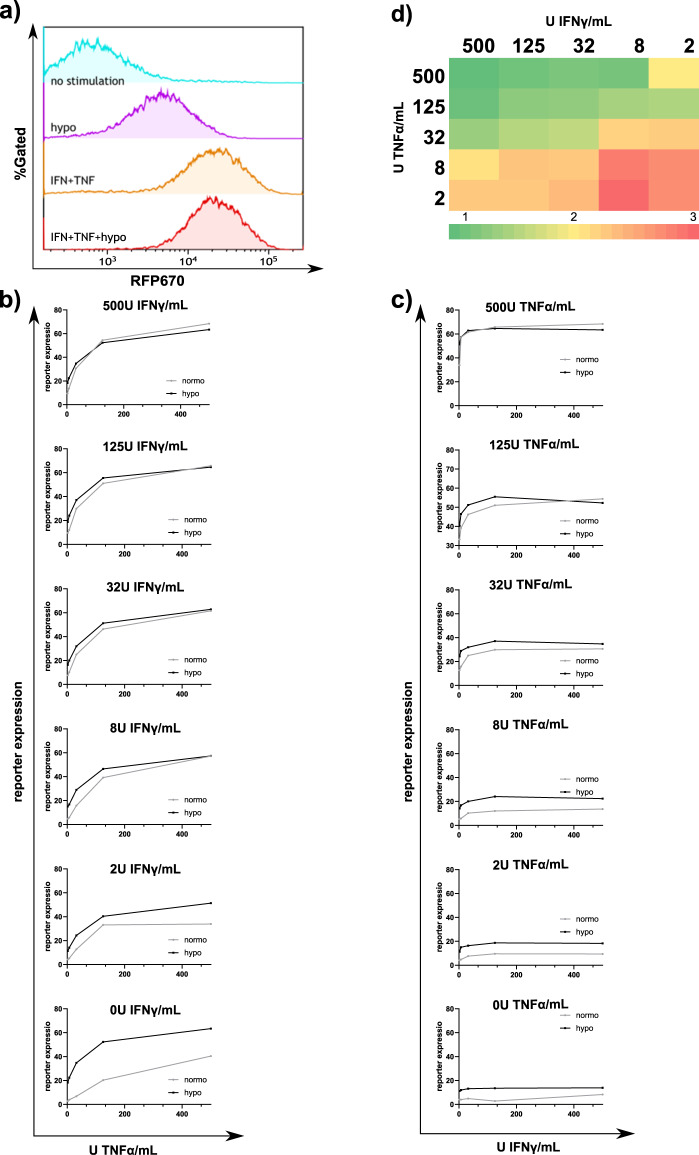
Fig. 4. The G1K06H1 promotor shows an additive response to hypoxia and cytokines with physiological cytokine concentrations.
a Representative plots of HEK293T cells infected using lenti viral vectors with RFP670 under the control of the G1K06H1 promotor and ZsGreen controlled by the ef1α core promotor. At 72 h following infection, the cells were treated for 48 h with 500 U/ml indicated cytokines and for 18 h under hypoxic or normoxic conditions, harvested and analyzed by flow cytometry. Data shown are ZsGreen-positive, single-discriminated and DAPI-negative results. b From the top panel to the bottom—cells were treated for 48 h with 500, 125, 32, 8, 2 or 0 U/mL of TNF respectively and veering IFN concentrations and placed under hypoxic or normoxic conditions for 18 h, cells were analyzed by flow cytometry as described above. Showing average of duplicates, error bars indicate standard deviation. c From the top panel to the bottom—cells were treated for 48 h with 500, 125, 32, 8, 2 or 0 U/mL of IFN respectively and veering TNF concentrations and placed under hypoxic or normoxic conditions for 18 h, cells were analyzed by flow cytometry as described above. Showing average of duplicates, error bars indicate standard deviation. d Ratios of reporter expression with different cytokine concentrations under hypoxic or normoxic conditions. Results are from one representative experiment of four performed.

